Service Detail

Fundus Photography

We photograph your retina so we can monitor your eye health over time.

Service Overview

What this service covers

Fundus photography captures high-resolution images of the back of your eye, providing a visual record that allows for accurate tracking of changes over time. Non-mydriatic retinal photography does not require dilating drops, making it a comfortable and convenient choice for routine screening of conditions like diabetic and hypertensive retinopathy. When a wider peripheral view is needed, dilated fundus photography offers a more comprehensive examination, ideal for assessing retinal tears, choroidal lesions, and post-treatment monitoring.

Fundus photography at Lead Opticals

Service Details

Detailed service breakdown

Detail 01

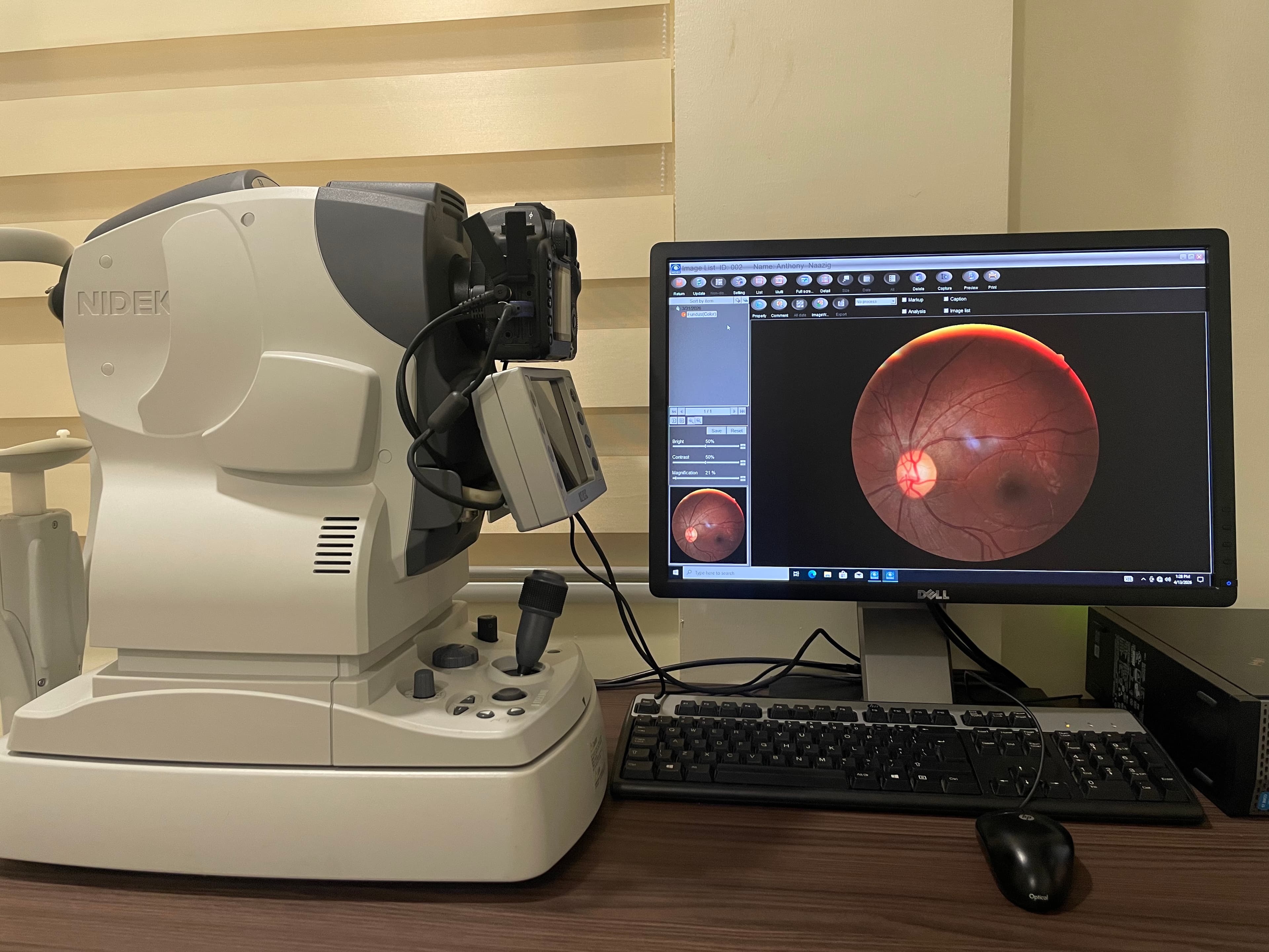
Non-Mydriatic Retinal Photography

High-resolution retinal images captured without dilating drops — fast and comfortable for the patient.

Includes

  • Optic disc documentation
  • Retinal baseline imaging
  • Diabetic retinopathy screening
  • Hypertensive retinopathy assessment

Clinical Note

Available without dilation in most patients — ideal for routine screening and annual comparison.

Detail 02

Dilated Fundus Photography

Wider-field retinal imaging following pupil dilation — allows a more complete peripheral view.

Includes

  • Peripheral retinal examination
  • Retinal tear and lattice degeneration
  • Post-treatment monitoring
  • Choroidal lesion documentation

Clinical Note

Dilation temporarily affects near vision and light sensitivity for 2 to 4 hours after the procedure.